...
GET IN TOUCH
Case Study

Reporter Cell Lines for Immuno-Oncology Drug Discovery: Targeting T-Cell Activation

Overview

Reporter cell lines are genetically engineered cells that express reporter genes, allowing monitoring of cellular processes through detectable signals like luminescence or fluorescence. In immuno-oncology, these cell lines are useful for monitoring T-cell activation, a critical functional readout for assessing immune responses to therapies. By linking reporter gene expression to pathways like the NFAT (Nuclear Factor of Activated T cells) pathway, these systems enable precise, quantifiable measurements of T-cell activation, critical for evaluating immunotherapies, offering valuable insights into the effectiveness of immunotherapies.

This case study details the development of a Jurkat-NFAT-Luc reporter cell line, optimized for sensitive, reproducible detection of T-cell activation. By incorporating an NFAT-responsive luciferase construct into the Jurkat T-cell background, this system offers a reliable platform for assessing immune-modulating agents, supporting early-stage evaluation and high-throughput screening.

Our Approach

We generated NFAT-Luc reporter Jurkat cells by introducing a plasmid carrying the luciferase gene under the NFAT response element via electroporation. Following antibiotic selection, stable Jurkat-NFAT-Luc cells were established. The functionality of these cells was validated by stimulating them with T-cell activators or chemical stimulants, to confirm stable integration and activation of luciferase expression through both TCR-dependent and independent signaling pathways.

Methodology

To evaluate the responsiveness of the Jurkat-NFAT-Luc reporter cell line to T-cell stimulation, a 96-well plate-based functional assay was performed that involved:

1. TCR-Dependent Stimulation Using Anti-CD3/CD28 Beads

  • Jurkat-NFAT-Luc and Jurkat wild-type (WT) cells (25,000 per well) were seeded in a 96-well culture plate containing complete medium (RPMI with 10% FBS) and incubated for 24 hours.
  • The next day, anti-CD3/CD28 activation beads were washed with phosphate-buffered saline (PBS) and resuspended in complete medium.
  • Beads were added to wells at varying cell-to-bead ratios (1:0 to 1:8).
  • Cells were incubated at 37°C with 5% CO2 for 6 hours.
  • After incubation, cells were lysed using a luciferase assay reagent.
  • Luminescence was measured using a multi-mode plate reader

2. TCR-Independent Stimulation Using PMA and Ionomycin
  • Jurkat-NFAT-Luc and Jurkat-WT cells (50,000 per well) were treated with a mixture of PMA (0.081 µM) and ionomycin (1.34 µM) in complete medium.
  • Cells were incubated for 6 hours at 37°C in 5% CO2.
  • Following stimulation, cells were lysed using a luciferase assay reagent.
  • Luminescence was recorded using a multi-mode plate reader.

Outcomes

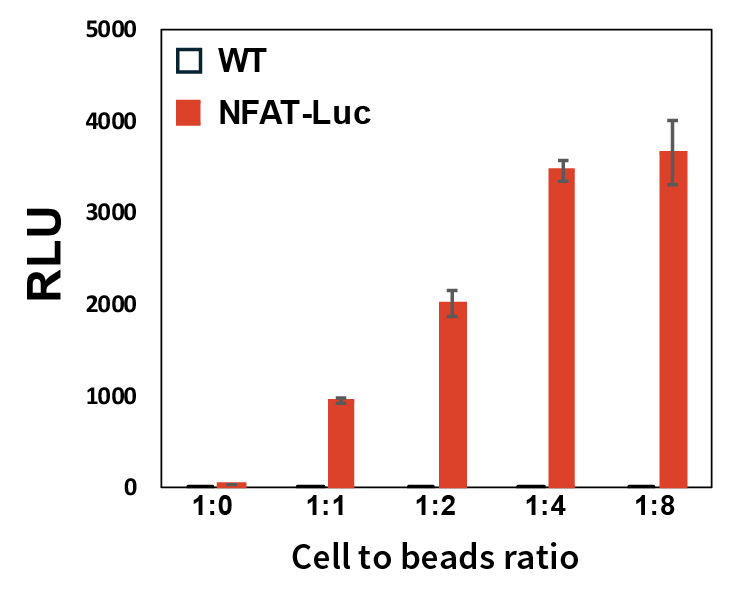
  • TCR-Dependent Stimulation with Anti-CD3/CD28 Beads: Jurkat-NFAT-Luc and Jurkat wild-type (WT) cells were stimulated with anti-CD3/CD28 beads at varying cell-to-bead ratios for 6 hours. This stimulation activates the TCR/CD3 complex and provides a costimulatory signal through CD28, mimicking T-cell activation via antigen-presenting cells. Activation of the TCR/CD3 pathway triggers the translocation of the NFAT transcription factor to the nucleus, inducing the expression of cytokines such as IFN-γ and TNF-α. The luciferase activity measured in the assays reflects T-cell activation via the NFAT pathway, demonstrating high specificity, precision, and linearity (Figure 1), and confirming the ability of the Jurkat-NFAT-Luc cell line to accurately monitor T-cell activation in response to immune-modulating agents.

Figure 1: Response of Jurkat-NFAT-Luc and Jurkat wild-type (WT) cells to anti- CD3/CD28 beads. Error bars represent ±SE.
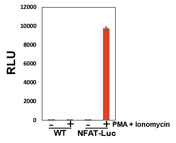
  • TCR-Independent Stimulation with PMA and Ionomycin: Jurkat-NFAT-Luc and Jurkat-WT cells were treated with a combination of PMA and ionomycin for 6 hours. This treatment bypasses the TCR/CD3 complex and stimulates the NFAT pathway through protein kinase C (PKC) activation and increased calcium influx. Consequently, NFAT translocates to the nucleus, leading to luciferase expression. A significant increase in the luciferase activity (Figure 2) confirms T-cell activation through a TCR-independent mechanism, demonstrating the Jurkat-NFAT-Luc cell line’s responsiveness to various signaling pathways and its consistency in activation assays.

Figure 2: Response of Jurkat-NFAT-Luc cell line upon treatment with a combination of PMA and ionomycin. Error bars represent ±SE.

These findings confirm the Jurkat-NFAT-Luc cell line as a reliable tool for tracking T-cell activation, both through TCR-dependent and TCR-independent pathways, supporting its use in evaluating immuno-oncology therapeutics.

Why Aragen?

At Aragen, we specialize in custom cell line development for drug discovery, including immuno-oncology applications.

We offer:

  • Comprehensive Solutions: A complete range of cell lines (CHO, HEK293, cancer cells) and reporter genes (GFP, luciferase).
  • Rigorous Quality Control: Extensive testing for expression levels, mycoplasma, sterility, and stability, ensuring reliable results.
  • Flexible Platforms: Choose from different cell backgrounds and expression systems (intracellular, secretory, membrane) designed for high-yield production and functional assays.
  • Immuno-Oncology Expertise: Specialized reporter cell lines like Jurkat-NFAT-Luc track T-cell activation, ideal for evaluating checkpoint inhibitors, CAR-T cells, and TCR modulators.
  • Efficient Timelines: Tool cell lines in 4-6 weeks, monoclones in 10-12 weeks, and production cell lines in 16-18 weeks, accelerating your research.

Accelerate your research with our reliable, high-quality cell line development. Visit our website and get in touch to learn more!

Seraphinite AcceleratorOptimized by Seraphinite Accelerator
Turns on site high speed to be attractive for people and search engines.